alarm RGCIRC, Niti Bagh alarm Emergency - 24/7 Book an Appointment Find a Doctor

Biopsy Procedure at RGCIRC

Biopsy Procedure at RGCIRC

A biopsy is one of the most important tests used to confirm or rule out cancer. When a lump, abnormal growth, suspicious lesion, or unusual imaging finding needs further evaluation, a biopsy helps doctors study the tissue closely and understand whether the cells are cancerous, non-cancerous, or require additional testing.

At RGCIRC (Rajiv Gandhi Cancer Institute and Research Centre), biopsy procedures are performed using a wide range of techniques, depending on the tumour’s location, size, accessibility, and the patient’s clinical condition. Every biopsy is supported by careful planning from the treating team, so that the right tissue sample is collected safely and accurately.

What happens after the tissue is collected is equally important. Biopsy samples at RGCIRC are processed through its NABL-accredited molecular laboratory, one of the first dedicated oncology molecular laboratories in India, where pathological, immunohistochemical, and molecular analysis can be performed. This integrated approach helps cancer specialists arrive at an accurate diagnosis and plan treatment based on the exact nature of the disease.

What is a Biopsy?

A biopsy is a medical procedure in which a small sample of tissue is removed from the body and examined under a microscope by a pathologist. The word comes from the Greek words “bios”, meaning life, and “opsis”, meaning sight. In simple terms, it means looking closely at living tissue.

In cancer diagnosis, a biopsy is often the most definitive way to confirm whether abnormal cells are cancerous or non-cancerous. While imaging scans, blood tests, tumour markers, and clinical examination can show that something may be suspicious, they usually cannot confirm the exact nature of the disease on their own.

A biopsy helps the pathologist identify the type of cells present, whether they are malignant, how abnormal they look, and how the disease is likely to behave. This information is important because different cancers can require very different treatment approaches.

In the era of precision oncology, biopsy testing has become even more important. At RGCIRC, biopsy samples can be evaluated not only for diagnosis but also for deeper tumour profiling, including immunohistochemistry and molecular testing when required. These tests may help identify mutations, gene changes, protein expression, and biomarkers that can guide treatment decisions.

This means a biopsy does more than confirm the presence of cancer. It helps doctors understand the cancer’s biology and decide whether chemotherapy, targeted therapy, immunotherapy, surgery, radiation therapy, or a combination of treatments may be appropriate for the patient.

Types of Biopsy

The right biopsy technique depends on several factors, including where the suspicious tissue is located, how deep it is, its size, and the type of information the clinical team needs. At RGCIRC, the biopsy method is selected after clinical assessment so that the most suitable and safe approach can be planned for each patient.

Fine Needle Aspiration Cytology (FNAC)

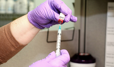
Fine Needle Aspiration Cytology, or FNAC, uses a thin needle to collect a small sample of cells from a lump or mass. It is a minimally invasive technique and is commonly used for accessible lumps in areas such as the neck, thyroid, breast, and lymph nodes.

FNAC helps doctors study individual cells under a microscope. In many cases, it may be performed with ultrasound guidance to improve accuracy.

Core Needle Biopsy

A core needle biopsy uses a slightly larger needle to remove a small cylinder of tissue. Unlike FNAC, which mainly collects cells, a core biopsy provides a tissue sample that shows how the cells are arranged within the tissue.

This makes it useful for many cancers, including breast, prostate, liver, and soft tissue tumours. Depending on the location of the lesion, the biopsy may be guided by ultrasound, CT, or MRI.

Image-Guided Biopsy

Some lesions are located deep inside the body and cannot be felt from the surface. In such cases, image-guided biopsy helps doctors reach the correct area more precisely.

This technique may use ultrasound, CT, MRI, or PET-CT guidance, depending on the location and nature of the suspicious area. Image-guided biopsies may be used for lung lesions, liver masses, abdominal or retroperitoneal tumours, bone lesions, and other deep-seated abnormalities.

Endoscopic Biopsy

An endoscopic biopsy is performed using a thin, flexible tube with a camera, called an endoscope. The scope is passed through a natural opening, such as the mouth or rectum, to reach the area of concern. Small instruments are then used to collect a tissue sample.

This type of biopsy may be used for conditions involving the oesophagus, stomach, colon, rectum, airways, or biliary system. Depending on the case, procedures such as upper GI endoscopy, colonoscopy, bronchoscopy, ERCP, or endoscopic ultrasound may be used.

Endobronchial Ultrasound-Guided Biopsy

Endobronchial ultrasound-guided biopsy, or EBUS, combines bronchoscopy with ultrasound imaging. It helps doctors collect samples from lymph nodes or masses near the airways in the chest.

EBUS is especially useful in lung cancer diagnosis and staging, as it can help evaluate mediastinal lymph nodes without the need for more invasive surgical procedures in selected patients.

Bone Marrow Biopsy

A bone marrow biopsy involves collecting a sample of bone marrow, usually from the back of the hip bone. It is commonly used to diagnose or assess blood cancers such as leukaemia, lymphoma, and multiple myeloma.

The test helps doctors understand how blood cells are being produced and whether cancer cells or abnormal blood cells are present in the marrow.

Excisional and Incisional Biopsy

An excisional biopsy removes the entire suspicious lesion, while an incisional biopsy removes only a part of it. These are surgical biopsy procedures and may be performed under local or general anaesthesia, depending on the location and size of the lesion.

An excisional biopsy may be used for small, accessible lesions. An incisional biopsy may be preferred when the lesion is larger or when removing the entire mass at the diagnostic stage is not appropriate. In suspected sarcoma cases, biopsy planning is especially important and should be done by the treating cancer team.

Punch Biopsy and Shave Biopsy

Punch biopsy and shave biopsy are commonly used for skin lesions. A punch biopsy uses a circular blade to remove a small, deeper sample of skin and underlying tissue. A shave biopsy removes the upper layers of a skin lesion.

The choice between these techniques depends on the appearance, depth, and clinical suspicion of the lesion.

Sentinel Lymph Node Biopsy

Sentinel lymph node biopsy is a procedure used to check whether cancer has spread to the first lymph node or group of lymph nodes that drain the tumour area. These are called sentinel lymph nodes.

If the sentinel lymph node does not show cancer spread, further lymph node removal may not be needed in selected cases. This can help reduce the risk of complications such as lymphoedema. Sentinel lymph node biopsy is commonly used in early breast cancer and may also be considered in selected head and neck, skin, and gynaecological cancers.

How is a Biopsy Performed?

The steps of a biopsy depend on the type of biopsy and the area of the body being tested. However, most biopsy procedures follow a similar process before, during, and after the test.

Before the Procedure

Before a biopsy, the clinical team reviews the patient’s symptoms, examination findings, imaging reports, and relevant blood tests. This helps them identify the exact area to be sampled and choose the safest and most suitable biopsy technique.

Patients may be asked about current medicines, especially blood thinners, as some medications may need to be paused before the procedure. For certain image-guided or endoscopic biopsies, fasting may be required for a few hours. The procedure, possible risks, and expected recovery are explained to the patient, and written consent is taken before the biopsy.

During the Procedure

Most biopsies are performed under local anaesthesia, which means the area is numbed while the patient remains awake. Sedation may be used for some endoscopic procedures, while general anaesthesia may be required for selected surgical biopsies.

The procedure may take around 15 to 45 minutes, depending on the biopsy type and the site being sampled. In image-guided biopsies, the doctor us es ultrasound, CT, MRI, or another imaging method to guide the needle accurately to the target area. Patients may feel pressure or mild discomfort, but pain is usually reduced with anaesthesia.

After the Procedure

After the biopsy, pressure is applied to the site to reduce bleeding, and a dressing is placed. Many patients can go home the same day after a short period of observation.

Some biopsies, such as lung, liver, or deep-seated image-guided biopsies, may require longer monitoring to check for complications. The care team provides specific instructions about rest, wound care, medicines, activity restrictions, and when to seek medical help.

Specimen Processing

After the tissue sample is collected, it is carefully labelled, preserved, and sent to the pathology department for analysis.

At RGCIRC, biopsy samples are evaluated using appropriate diagnostic methods, which may include routine microscopy, special stains, immunohistochemistry, biomarker testing, and molecular testing such as next-generation sequencing when clinically indicated. This helps the cancer team understand the exact nature of the disease and plan treatment more accurately.

Understanding Your Biopsy Report

A biopsy report is a pathology report that explains what was found in the tissue sample and what it may mean for diagnosis and treatment. While your doctor will explain the report in detail, understanding the main terms can help you follow the discussion more clearly.

A biopsy report may include:

  • Histological diagnosis: This confirms whether cancer cells are present and identifies the type of cancer, such as adenocarcinoma, squamous cell carcinoma, lymphoma, or another diagnosis.
  • Tumour grade: This describes how abnormal the cancer cells look compared with normal cells. In general, higher-grade cancers may behave more aggressively, but the meaning of the grade depends on the cancer type.
  • Margins: In excisional biopsies or surgical specimens, the report may mention whether the edges of the removed tissue are clear or involved. Clear margins mean no cancer cells are seen at the outer edge of the tissue sample. Involved margins may mean that further treatment or surgery needs to be considered.
  • Immunohistochemistry: This test looks for specific proteins in the cancer cells. It may help confirm the cancer type and guide treatment decisions. Examples include hormone receptors, HER2 status, PD-L1 expression, and other markers, depending on the cancer.
  • Molecular testing: When advised, molecular testing looks for gene mutations, fusions, amplifications, or other biomarkers. These results can help doctors decide whether targeted therapy, immunotherapy, chemotherapy, or another treatment approach may be suitable.

How Long Does the Report Take?

The time taken for a biopsy report can vary depending on the type of sample and the tests required. A standard histopathology report may be available within a few working days. If additional tests such as immunohistochemistry, biomarker testing, or molecular testing are needed, the report may take longer.

For second opinion requests on biopsy material sent from other hospitals, RGCIRC states that the usual turnaround time for the report is 4 to 5 working days.

Is Biopsy Painful?

This is one of the most common questions patients have before a biopsy, and it deserves a clear answer.

Most biopsies cause only mild discomfort because local anaesthesia is used to numb the area before the sample is taken. During the procedure, patients may feel pressure, pulling, or a brief sharp sensation, but usually not severe pain.

Some biopsy procedures, such as bone marrow biopsy, may cause a deeper ache or more noticeable discomfort, even with local anaesthesia. In selected cases, sedation may be considered to help the patient stay more comfortable.

After the procedure, mild soreness, bruising, or tenderness at the biopsy site is common for 24 to 48 hours. This usually improves with rest and simple pain relief advised by the doctor. Most patients can return to normal daily activities within a day or two after a needle biopsy. Recovery may take a few days to a week after a surgical biopsy, depending on the site and procedure.

For many patients, the anxiety before a biopsy is greater than the physical discomfort of the procedure itself. At RGCIRC, the clinical team explains what to expect, answers questions, and takes steps to keep patients as comfortable as possible throughout the biopsy process.

Biopsy at RGCIRC

At RGCIRC, biopsy procedures are planned according to the patient’s condition, the suspected cancer type, and the location of the abnormal tissue. The procedure is performed by the specialist best suited for that biopsy technique, such as an interventional radiologist for image-guided biopsies, a pulmonologist for bronchoscopic or EBUS-guided biopsies, a surgeon for excisional or sentinel lymph node biopsies, or a haematologist for bone marrow biopsies.

This helps ensure that the biopsy is not only technically accurate, but also aligned with the overall cancer treatment plan.

NABL-Accredited Molecular Laboratory

A biopsy does not end when the tissue sample is collected. What happens to the sample after collection is equally important for diagnosis and treatment planning.

RGCIRC’s molecular diagnostics laboratory is NABL-accredited and was developed under the leadership of Dr Anurag Mehta. The laboratory offers more than 100 tests across infectious disease, genetic, and oncology testing. Its testing platforms include real-time PCR, Fluorescent In Situ Hybridisation, Pyrosequencing, Sanger sequencing, and high-throughput Next Generation Sequencing for genetic predisposition testing, tumour-specific panels, tumour mutation burden, and liquid biopsy.

This diagnostic depth helps the clinical team move beyond confirming or ruling out cancer. Where indicated, biopsy samples can provide important molecular information that may guide precision treatment decisions, including targeted therapy, immunotherapy, chemotherapy, or other treatment approaches.

Second Opinion in Pathology

Patients who have already had a biopsy elsewhere can also request a second opinion in pathology at RGCIRC. Histopathology or cytopathology biopsy material, including slides, blocks, or specimens, may be submitted along with the previous biopsy report, relevant medical records, imaging reports, and treatment details.

RGCIRC’s pathology team reviews the biopsy material and may perform routine microscopy, special stains, immunohistochemistry, biomarker testing, theranostic marker studies, and molecular tests when required. According to RGCIRC, the usual turnaround time for pathology second opinion reports is 4 to 5 working days. Reports are sent to the registered email ID or can be collected from the OPD report dispatch counter.

This service gives patients and families an opportunity to confirm the diagnosis, understand the disease more clearly, and make more informed treatment decisions.

Why Choose RGCIRC for Biopsy?

Choosing where to have a biopsy is important because the biopsy often becomes the first step in diagnosis, staging, and treatment planning. The accuracy of the procedure, the quality of tissue processing, and the expertise used to interpret the report can all influence what happens next.

At RGCIRC, biopsy services are supported by specialist teams, advanced imaging guidance, oncology-focused pathology, molecular diagnostics, and multidisciplinary cancer care.

Comprehensive Biopsy Services Under One Roof

RGCIRC offers a wide range of biopsy techniques for different cancer types and body sites. These may include FNAC, core needle biopsy, CT-guided biopsy, ultrasound-guided biopsy, PET-guided biopsy, bronchoscopy-guided biopsy, EBUS-guided biopsy, endoscopic biopsy, bone marrow biopsy, sentinel lymph node biopsy, skin biopsy, and surgical biopsy.

The appropriate technique is chosen based on the patient’s condition, the location of the suspicious tissue, and the clinical information needed for diagnosis and treatment planning. This integrated setup helps patients receive diagnostic evaluation within a comprehensive oncology centre.

Expertise in Interventional Oncology

RGCIRC’s Interventional Oncology service supports image-guided procedures for deep-seated or difficult-to-access lesions. These procedures may be performed using CT, ultrasound, or other imaging guidance to help doctors reach the target area accurately.

RGCIRC states that it was the first hospital in India to establish a dedicated Interventional Oncology service line. Its interventional oncology team performs procedures in a state-of-the-art oncology Cath Lab or under CT/USG guidance, including complex biopsies that help provide tissue for histopathological diagnosis and mutational analysis.

Molecular Diagnostics for Precision Oncology

At RGCIRC, biopsy samples are supported by oncology-focused pathology and molecular diagnostics. Its NABL-accredited molecular laboratory offers more than 100 tests across infectious disease, genetic, and oncology testing.

The laboratory uses platforms such as real-time PCR, Fluorescent In Situ Hybridisation, Pyrosequencing, Sanger sequencing, and high-throughput Next Generation Sequencing. These tests may help identify biomarkers, gene changes, tumour mutation burden, and other molecular details that can guide treatment planning.

This means the biopsy report may do more than confirm whether cancer is present. Where indicated, it can help doctors understand the biology of the tumour and plan treatment more precisely.

Multidisciplinary Review of Biopsy Results

At RGCIRC, biopsy findings are considered along with clinical examination, imaging, staging, and the patient’s overall condition. The institute follows an organ-specific multidisciplinary approach to cancer diagnosis and treatment, with tumour board review for more complex cases.

This helps ensure that treatment decisions are not based on the pathology report alone, but on the complete clinical picture.

Accredited Diagnostic and Hospital Standards

RGCIRC is accredited by NABH and NABL and also has Green OT and Nursing Excellence certifications. These accreditations reflect recognised standards in hospital care, laboratory quality, patient safety, and clinical processes.

The institute has also been recognised among the World’s Best Specialised Hospitals by Newsweek in 2024 and 2025, ranked No. 1 in North India among single-specialty hospitals in The Times of India’s 2025 survey, and awarded Best Oncology Hospital of the Year 2026 at the 7th Edition of the Cancer Summit Awards organised by the IHW Council.

Commitment to Accessible Cancer Care

RGCIRC is a not-for-profit institution established with quality, affordability, and access as key goals. Since its inception in 1996, the institute has touched the lives of more than 3.5 lakh patients from India, neighbouring SAARC countries, and other regions.

Through its philanthropic initiatives, RGCIRC works to reduce financial barriers for eligible patients and support wider access to cancer diagnosis and treatment.

Book a Biopsy Consultation at RGCIRC

If you have been advised to undergo a biopsy, or if you already have biopsy slides, blocks, or reports from another hospital and want them reviewed, you can consult the specialist team at RGCIRC.

The team can help you understand whether a biopsy is needed, which biopsy technique may be suitable, and whether additional pathology, immunohistochemistry, biomarker, or molecular testing may be required for diagnosis and treatment planning.

To book a consultation or access biopsy-related services at RGCIRC:
Book online: care.rgcirc.org
Call Rohini: +91-11-4702 2222
Call Niti Bagh, South Delhi: +91-11-4582 2222
OPD Hours: Monday to Saturday, 9:00 AM to 5:00 PM
Emergency Services: 24×7 at both campuses

Frequently Asked Questions

What is a biopsy test and why is it done?

A biopsy is a procedure in which a small sample of tissue or cells is removed from the body and examined in a laboratory. It is done to confirm or rule out cancer, identify the cancer type, assess how the disease may behave, and guide treatment planning. In many cases, a biopsy is the most definitive way to confirm a cancer diagnosis.

What are the different types of biopsy?

Common biopsy types include Fine Needle Aspiration Cytology (FNAC), core needle biopsy, image-guided biopsy, endoscopic biopsy, bronchoscopy-guided biopsy, EBUS-guided biopsy, bone marrow biopsy, sentinel lymph node biopsy, excisional biopsy, incisional biopsy, punch biopsy, and shave biopsy. The right type depends on the location, depth, size, and nature of the suspicious tissue.

Is a biopsy painful?

Most biopsies cause only mild discomfort because local anaesthesia is used to numb the area. Patients may feel pressure, pulling, or brief discomfort, but usually not severe pain. Mild soreness, bruising, or tenderness may occur for one to two days after the procedure. Bone marrow biopsy can feel more uncomfortable, and sedation may be considered in selected cases.

How long does a biopsy report take?

The time taken for a biopsy report depends on the sample type and the tests required. A standard histopathology report may be available within a few working days. If immunohistochemistry, biomarker testing, or molecular testing is needed, the report may take longer. For second opinion pathology reviews on biopsy material sent from another hospital, RGCIRC states that the usual turnaround time is 4 to 5 working days.

What does a biopsy report tell you?

A biopsy report may confirm whether cancer is present and identify the specific cancer type. It may also mention the tumour grade, margin status in surgical specimens, immunohistochemistry results, and molecular testing findings where required. These details help doctors understand the disease better and plan treatment more accurately.

Can I get a second opinion on my biopsy at RGCIRC?

Yes. RGCIRC offers second opinion in pathology for biopsy material processed at another hospital. Patients can submit histopathology or cytopathology material such as slides, blocks, or specimens for review. The review may include routine microscopy, special stains, immunohistochemistry, biomarker testing, theranostic marker studies, and molecular testing when required.

What is a core needle biopsy?

A core needle biopsy uses a hollow needle to remove a small cylinder of tissue from a suspicious area. It provides more information than FNAC because it shows the tissue structure, not just individual cells. This can help doctors identify the cancer type, grade, and other features needed for treatment planning.

What is the cost of a biopsy test in Delhi?

The cost of a biopsy test in Delhi depends on the type of biopsy, the body site, whether image guidance is needed, and the laboratory tests required after the sample is collected. At RGCIRC, a cost estimate is usually provided after consultation and clinical assessment. Eligible patients who need financial support may also enquire about assistance through RGCIRC’s philanthropic services.

Patient & Family