

Basis of PET-CT Scan
PET-CT
It is a state-of-the-art technology that has changed the oncology practice in the last two decades. It helps in practicing precise and personalized medicine. Current PET equipment is an integrated PET-CT machine that allows the acquisition of both PET and CT on one platform. PET provides the essential signals of the tumor, and CT helps in localization. Upon requiring a regional contrast-enhanced CT (CECT) scan, it can also be performed with the same equipment in the same sitting. PET-CT is helpful in early diagnosis, response assessment, restaging, and follow-up of cancer patients. Many a time, it helps in identifying the correct site of tumor biopsy and reduces the multiple procedures.
We installed the First PET-CT scanner in January 2008, and this was a Hi-Rez Tru point Siemens system integrated into a 40 slice CT scanner. The second PET-CT scanner was installed in November 2015, and that was a ‘Molecular PET-CT’ (mCT) system by Siemens. This equipment was unique because it has an extra ring of high sensitive PET detector, named as Tru V system, which allows faster imaging with lesser radiation dose. With the commitment to provide the most updated technology, we have replaced our first PET-CT scanner with the most advanced system, a ‘Digital PET-CT’ in November 2019. With this novel, cutting-edge technology, we provide a new horizon to our patients beyond analog technology.
Radiopharmaceuticals used in PET-CT
Positron emission tomography (PET) is an advanced diagnostic imaging technique that is used in various cancers at multiple stages of their management. This technology exploits many biological pathways or antigens, enhanced or over expressed in the tumor cells. A suitable ligand labelled with a positron-emitting radionuclide is used to target this antigen or biological pathway. Many PET-CT radiopharmaceuticals for targeting different types of cancers are available, e.g., FDG, PSMA, DOTANOC, DOTATATE, DOPA, FAPI, FES, MESO, etc.
Conventional 18F-FDG PET-CT Scan
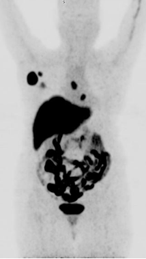
2-[fluorine18]-fluoro-2-deoxy-D-glucose (FDG) is the key radiopharmaceutical PET imaging. FDG is an analogue of the glucose. Like glucose, it is transported into the tumor cells, by means of glucose transporter protein (GLUT receptors) and subsequently it is phosphorylated by an enzyme hexokinase to FDG-6-phosphate. As FDG-6-phosphate is not a substrate for glucose-6-phosphate isomerase enzyme (the next step in glycolysis), it is biochemically trapped within the cell. This process of high FDG uptake and trapping in tumor cell constitutes the basis for imaging of distribution of tracer with PET. Since, there is many fold increase in glucose metabolism in malignant tumors as compared to normal, it is easy to detect this difference in metabolism using PET. Therefore, PET has high sensitivity and high negative predictive value as compared to conventional morphological modalities like computed tomography (CT), ultrasonography (USG), magnetic resonance imaging (MRI), which use size / density changes as the only or major criterion to distinguish between benign and malignant diseases. FDG PET has a well-established role in initial staging, restaging and monitoring the response to the therapy in the management of various cancers.
Fusion of PET & CT allows precise localization of the diseased sites, for optimal management of patient with cancer. Co-registration of a structural and functional image provided by PET-CT is essential for accurate planning of surgical procedures. Likewise, PET-CT imaging is becoming a standard of care in Radiation Oncology. PET guided IMRT can selectively target head and neck cancers, while reducing critical normal tissues. Staging, early response evaluation, detection of recurrence and follow-up of most of the malignancies can be done with this modality. A PET guided biopsy from the most metabolically active site gives the best chance to identify the tumor and reduced futile attempts.
We have standardized side by side fusion of PET-MRI images, which provide excellent soft tissue delineation with metabolic parameters, and are now being routinely used in lung, Head & Neck cancers & also in Gynaecological Malignancies.
Who Needs PET CT Scan?
- For detection and staging of disease
- For early evaluation of response, differentiate between responder & non responder
- Effectiveness of treatment
- Detection of early recurrence
- RT planning
- PET guided biopsy
Specialized PET CT Radiopharmaceuticals
68Ga-DOTANOC PET-CT
This is the standard PET used in well differentiated neuroendocrine tumours (WHO grade 1 or 2). These tumor cells over expressed SSTR receptors 2,3,5. 68Ga-DOTANOC or DOTATATE both can be used to target these SSTR receptors. DOTANOC PET is used for staging, treatment response assessment, suspected recurrence and restaging. DOTANOC PET is also useful in medullary thyroid carcinoma (MTC) and pheochromocytoma, paraganglioma and neuroblastoma.
68Ga-PSMA PET-CT
Prostate specific membrane antigen (PSMA) is a specific PET tracer used in prostate cancer with following indications.
- For detection and staging of disease
- For evaluation of response, differentiate between responder & non responder
- Effectiveness of treatment
- Detection of early recurrence
- RT planning
- PET guided biopsy
18F-DOPA PET-CT
18F-fluoro-3,4-dihydroxyphenylalnine (18F-DOPA) is a PET tracer that measures the uptake of dopamine precursors for assessment of presynaptic dopaminergic integrity. It is the standard PET used in Parkinson’s disease (PD). In oncology, it is the tracer of choice in suspected medullary thyroid carcinoma (MTC) and high grade gliomas recurrence.
18F-FES PET-CT
18F-Fluoroestradiol (FES) PET is useful in estrogen receptor (ER) positive breast cancer. It is recently FDA approved for recurrent or metastatic breast cancer patients as an adjunct to biopsy. Establishing ER positive status in each metastatic breast cancer sites will help in selecting the appropriate and effective treatment.
18F-FES PET scan showing ER receptor positive right breast lesion with right axilla and mediastinal lymph nodes