
alarm RGCIRC, Niti Bagh alarm Emergency - 24/7 Book an Appointment Find a Doctor

RGCIRC Team

Cancer

26 October, 2026

Medical imaging plays an important role in cancer care, often helping doctors find answers that symptoms and physical examinations alone cannot provide. From identifying a suspicious growth to understanding how far cancer has spread and whether treatment is working, imaging tests such as X-rays, ultrasound, CT, MRI and PET-CT support medical decisions at different stages of a patient’s journey.

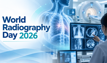
Making these images useful for patient care requires more than advanced technology. It also depends on trained professionals who perform imaging examinations safely, ensure image quality and help clinical teams obtain the information they need. World Radiography Day 2026, recognises the contribution of radiography professionals and highlights the role medical imaging continues to play in modern cancer diagnosis and treatment.

Key Takeaways

  • World Radiography Day is observed on 8 November every year, marking the anniversary of Wilhelm Conrad Röntgen’s discovery of X-radiation in 1895.
  • Medical imaging supports many stages of cancer care, from detecting an abnormality and guiding a biopsy to staging cancer, planning treatment and monitoring response.
  • Different imaging tests answer different clinical questions. A patient does not necessarily need every type of scan.
  • X-ray and CT use ionising radiation, while MRI and ultrasound do not.
  • Radiographers, radiologists, nuclear medicine professionals and other specialists work together with oncology teams to obtain and use imaging safely and effectively.
  • At RGCIRC, radiology, nuclear medicine and interventional oncology are integrated into multidisciplinary cancer care.

What is World Radiography Day and Why is it Celebrated on 8 November?

World Radiography Day is observed every year on 8 November to recognise the contribution of radiography to healthcare and increase awareness of diagnostic imaging and radiation therapy.

The date marks Wilhelm Conrad Röntgen’s discovery of X-radiation on 8 November 1895, a breakthrough that made it possible to see structures inside the human body without surgery.

Since then, medical imaging has advanced far beyond conventional X-rays. Technologies such as CT, MRI, ultrasound, mammography and PET-CT now help healthcare teams investigate disease, guide treatment and monitor patients across different stages of care. The day also recognises radiography as a profession and the role radiography professionals play in delivering safe and effective imaging.

8 November is also observed as the International Day of Radiology (IDoR), which focuses more broadly on the contribution of radiology to patient care. In 2026, the official IDoR campaign highlights how radiology can support earlier detection, reduce the need for some surgical procedures and contribute to better treatment outcomes.

How Does Medical Imaging Support Cancer Care?

Cancer care often involves answering a series of questions: Where is the abnormality? What is it likely to be? Has it spread? Which treatment may be appropriate? Is the treatment working?

Medical imaging helps answer these questions at different stages of care:

  • Detecting suspicious changes: Imaging can reveal a mass, enlarged lymph node or other abnormality that may need further evaluation.
  • Supporting diagnosis: Scans help doctors assess the location, size and characteristics of an abnormality and decide what further tests may be needed.
  • Guiding biopsy: Ultrasound, CT, mammography or other imaging techniques can help doctors accurately guide a needle to the area from which a tissue sample is required.
  • Staging cancer: CT, MRI, PET-CT and other imaging tests can help assess the tumour, nearby structures and lymph nodes, and look for signs of spread to other parts of the body.
  • Planning treatment: Imaging helps surgeons plan procedures and supports radiation oncology teams in defining the area that needs treatment while limiting exposure to surrounding healthy tissues.
  • Assessing treatment response: Comparing images taken before, during or after treatment can help doctors see how the cancer is responding.
  • Supporting follow-up: Imaging may be used after treatment when there are symptoms, examination findings or other concerns that need further evaluation.

Medical imaging therefore supports much more than cancer detection. It can provide important information from the initial evaluation through treatment planning, response assessment and follow-up.

Imaging and Image-Guided Cancer Care at RGCIRC

At Rajiv Gandhi Cancer Institute & Research Centre (RGCIRC), radiology is integrated with oncology care so that imaging findings can contribute directly to multidisciplinary decision-making and tumour board discussions.

Different imaging technologies are used according to the type of cancer, body area being examined and clinical question that needs to be answered.

X-Ray and Fluoroscopy

X-rays use a small amount of ionising radiation to create images of structures inside the body. In cancer care, they may be used for selected chest, bone or other examinations depending on the clinical need.

Fluoroscopy also uses X-rays, but instead of producing a single still image, it creates real-time moving images. This allows doctors to watch internal structures as they move and can also help guide certain diagnostic or interventional procedures.

At RGCIRC, digital X-ray and digital fluoroscopy are available as part of the institute’s imaging services, with images stored digitally so they can be reviewed and shared with the treating team as needed.

Ultrasound

Ultrasound uses high-frequency sound waves to create images of organs and soft tissues without using ionising radiation. In cancer care, it can help doctors examine areas such as the liver, abdomen, lymph nodes and other soft tissues, depending on the suspected condition.

Ultrasound can also be used to guide a needle accurately during certain procedures.

At RGCIRC, ultrasound and colour Doppler are used for diagnostic evaluation as well as image-guided procedures such as biopsy and fine-needle aspiration cytology (FNAC).

CT Scan

Computed tomography (CT) uses X-rays to create detailed cross-sectional images of the body. These images allow doctors to examine a tumour and the surrounding tissues in greater detail than a standard X-ray.

In cancer care, CT may be used to:

  • Assess the size and location of a tumour
  • Check nearby organs and lymph nodes
  • Help determine whether cancer has spread
  • Guide biopsies and other procedures
  • Support treatment planning

At RGCIRC, dual-energy CT imaging is available for diagnostic assessment, along with a dedicated CT scanner for interventional oncology procedures such as image-guided biopsies, complex drainage procedures and tumour ablation.

MRI

Magnetic resonance imaging (MRI) uses a strong magnetic field and radio waves to create detailed images of the body without ionising radiation.

MRI is particularly useful when doctors need a clearer view of soft tissues. Depending on the type of cancer, it may help assess areas such as the brain, spine, pelvis, liver and breast, as well as the relationship of a tumour to nearby structures.

At RGCIRC, 3 Tesla and 1.5 Tesla MRI systems with dedicated oncology protocols are used for detailed tumour assessment and, where appropriate, to help evaluate response to treatment.

Mammography and Breast Imaging

Mammography is a specialised low-dose X-ray examination of the breast. It is commonly used for breast cancer screening and can also help investigate symptoms or abnormalities detected during a clinical examination or another imaging test.

At RGCIRC, breast imaging services include full-field digital mammography and digital breast tomosynthesis, which creates multiple images of the breast from different angles. Image-guided procedures, including stereotactic and vacuum-assisted biopsy, are also available when further evaluation of a suspicious area is required.

PET-CT

Positron emission tomography-computed tomography (PET-CT) combines two types of information in one examination: PET shows areas of increased metabolic activity, while CT provides detailed anatomical images.

In selected cancers, PET-CT may help doctors:

  • Determine the extent of disease
  • Restage cancer after treatment or disease progression
  • Assess how well treatment is working
  • Investigate suspected recurrence
  • Support treatment planning
  • Identify a suitable area for biopsy in certain cases

At RGCIRC, PET-CT is used as part of cancer staging, restaging, treatment-response assessment and follow-up when clinically appropriate.

Imaging Can Also Guide Cancer Treatment

Modern radiology does more than produce diagnostic images.

Interventional oncology uses imaging to guide minimally invasive procedures inside the body. Instead of relying on a large surgical incision, specialists can use CT, ultrasound, fluoroscopy or catheter-based imaging to guide needles, catheters or treatment devices to a precise target.

At RGCIRC, image-guided procedures include:

  • CT- and ultrasound-guided biopsies
  • Drainage procedures
  • Radiofrequency, microwave and cryoablation for selected tumours
  • Embolisation procedures
  • Transarterial chemoembolisation (TACE) and radioembolisation (TARE) in selected cases
  • Image-guided procedures for cancer-related pain and other complications

RGCIRC has a dedicated Interventional Oncology service, where selected cancers and cancer-related complications can be treated using image guidance.

That said, not every patient needs every type of scan or image-guided procedure. The choice depends on the type and stage of cancer, previous test results and the specific information the treating team needs.

Who are the Professionals Behind Medical Imaging?

A scan may appear to be mainly about the machine, but producing useful medical images requires trained healthcare professionals working together.

Radiographers

Radiographers are healthcare professionals who perform imaging examinations and work closely with patients throughout the procedure.

Their responsibilities can include:

  • Preparing the patient for the examination
  • Correctly positioning the patient
  • Operating imaging equipment
  • Following the required imaging protocol
  • Applying radiation-protection measures where relevant
  • Checking that the images obtained are suitable for clinical use
  • Supporting patients who may be anxious, uncomfortable or unable to remain in a particular position

Radiography also includes therapeutic radiography, where trained professionals contribute to the planning and delivery of radiation therapy. Diagnostic and therapeutic radiographers therefore play important roles across screening, diagnosis, treatment and disease monitoring.

Radiologists

A radiologist is a medical doctor who specialises in interpreting medical images. They review scans alongside the patient’s symptoms, medical history and other test results to help identify abnormalities, assess disease and guide clinical decisions.

Some radiologists develop additional expertise in areas such as breast imaging, neuroradiology and cancer imaging, depending on the type of examination and condition being assessed.

Interventional Radiologists

Interventional radiologists are doctors who use medical imaging to guide minimally invasive procedures inside the body. Depending on the patient’s condition, these may include image-guided biopsies, drainage procedures, embolisation or tumour ablation.

Nuclear Medicine Professionals

Nuclear medicine professionals use small amounts of radioactive substances, called radiopharmaceuticals, to study how organs and tissues are functioning. In cancer care, techniques such as PET imaging can help assess disease activity, stage certain cancers and evaluate treatment response.

These imaging professionals work closely with pathologists, surgeons, medical oncologists and radiation oncologists. Bringing imaging findings together with pathology and other clinical information helps the treating team build a more complete picture of the cancer and plan the next steps in care.

How is Patient Safety Maintained During Medical Imaging?

It is common for patients to wonder whether imaging tests are safe, especially when radiation is involved. The answer depends on the type of examination being performed.

X-rays, CT scans, fluoroscopy and nuclear medicine procedures use ionising radiation, while MRI and ultrasound do not.

When ionising radiation is required, imaging teams aim to use the lowest amount needed to obtain useful diagnostic information. The examination should also have a clear medical reason so that the expected benefit outweighs the potential risk.

Safety checks go beyond radiation exposure. Before certain scans or procedures, the imaging team may ask about:

  • Pregnancy, where relevant
  • Kidney function before some contrast agents are given
  • Allergies or previous reactions to contrast material
  • Metal implants or medical devices before MRI
  • Blood sugar levels and preparation requirements before certain PET-CT studies
  • Medicines or medical conditions that may affect the examination
  • Previous scans, which may help avoid unnecessary repeat imaging

Patients should therefore share relevant medical conditions, pregnancy status, implants, allergies, medicines and any previous reactions with the imaging team before the test. These checks help the team choose the safest and most appropriate way to perform the examination.

From an X-Ray Discovery to Precision Cancer Care

When Röntgen discovered X-rays in 1895, medical imaging began with the ability to look inside the body without an operation. Today, imaging can do far more. It can show detailed anatomy, reveal metabolic activity, guide a biopsy needle to a specific lesion, help plan cancer treatment and measure how a tumour responds.

World Radiography Day is therefore not only about celebrating a scientific discovery. It is also about recognising the radiographers, radiologists, interventional specialists, nuclear medicine professionals and support teams who turn images into information that can guide patient care.

At RGCIRC, radiology, nuclear medicine and interventional oncology work alongside surgical, medical and radiation oncology teams to support coordinated cancer diagnosis, treatment planning and follow-up.

If you need an oncology consultation or further evaluation at RGCIRC, call +91-11-47022222 to book an appointment.

Frequently Asked Questions

1. Can a scan alone confirm that someone has cancer?

Not always. Imaging can identify an abnormality and provide important information about its location and characteristics, but many cancers require a biopsy and pathological examination for confirmation. The tests required depend on the suspected cancer and clinical findings.

2. Why might my doctor recommend more than one type of scan?

Different imaging tests provide different types of information. For example, CT may show anatomy and disease extent well, while MRI may provide more detailed soft-tissue information in certain areas. PET-CT can add information about metabolic activity. Doctors choose tests according to the specific clinical question rather than routinely ordering every available scan.

3. Does PET-CT detect every type of cancer?

No. PET-CT is useful for many cancers, but its value varies by cancer type, tumour biology and clinical situation. Some tumours may not show significant uptake with a particular PET tracer, so another imaging test may be more appropriate.

4. Should I take my previous scans when I go for a new imaging test?

Yes, when available. Previous images and reports can help radiologists compare changes over time and may provide important context for interpreting a new examination. RGCIRC can integrate outside imaging studies with in-house studies for review.

5. What is the difference between radiography and radiology?

Radiography refers to the professional practice involved in producing medical images and, in therapeutic radiography, contributing to radiation treatment. Radiology is the medical speciality in which doctors interpret imaging and may perform image-guided procedures. Radiographers and radiologists work closely together but have different professional roles.

6. Does RGCIRC perform image-guided biopsies?

Yes. At RGCIRC, biopsies can be performed under CT, ultrasound, mammography or MRI guidance, depending on the location and type of abnormality being investigated.

7. Can imaging show whether cancer treatment is working?

Yes. Depending on the cancer and treatment, follow-up CT, MRI, PET-CT or other imaging may be used to compare the tumour with earlier scans and assess treatment response. RGCIRC’s radiology services include treatment-response evaluation, while PET-CT is used in selected cancers for response assessment and restaging.

8. Does RGCIRC offer interventional oncology procedures?

Yes. RGCIRC provides interventional oncology procedures using CT, ultrasound or catheter-based image guidance. These include selected tumour ablation, embolisation, image-guided biopsy and other minimally invasive procedures. The appropriate procedure depends on the cancer type, location, disease extent and individual treatment plan.